
Enhanced tumorigenicity of CD133+EpCAM+ cells in NOD/SCID mice
To analysis the tumor initiating capability, NOD/SCID mice were transplanted with various amounts of CD133-EpCAM-, CD133+EpCAM-, CD133-EpCAM+ and CD133+EpCAM+ cells. In 10,000 cells group, CD133+EpCAM+ cells possessed higher tumorigenicity and faster tumor growth than other three phenotypes. In 5,000 and 1,000 cells groups, CD133+EpCAM+ cells possessed higher tumorigenicity and faster tumor growth compared to CD133-EpCAM+ and CD133-EpCAM- cells. In 500 cells group, only CD133+EpCAM+ cells formed tumor mass (Table 1, Fig. 6A). For example, CD133+EpCAM+ cells, but not CD133-EpCAM- cells, could efficiently initiate tumors in NOD/SCID mice (Fig. 6B). Sorted CD133+EpCAM+ cells formed similar histological features of xenograft tumors as unsorted Huh7 cells (Fig. 6C). The results showed that CD133+EpCAM+ cells embodied the increased tumorigenicity in vivo.
Table 1
Tumorigenicity of various Huh7 phenotypes in NOD/SCID mice.
| Phenotypes | Injecting numbers | Tumor incidencea |
|---|---|---|
| CD133+EpCAM+ | 10,000 | 6/7 |
| 5,000 | 4/6 | |
| 1,000 | 5/7 | |
| 500 | 2/6 | |
| CD133+EpCAM- | 10,000 | 2/6 |
| 5,000 | 2/7 | |
| 1,000 | 2/7 | |
| 500 | 0/7 | |
| CD133-EpCAM+ | 10,000 | 3/6 |
| 5,000 | 1/7 | |
| 1,000 | 0/7 | |
| 500 | 0/7 | |
| CD133-EpCAM- | 10,000 | 2/7 |
| 5,000 | 1/6 | |
| 1,000 | 0/7 | |
| 500 | 0/6 |
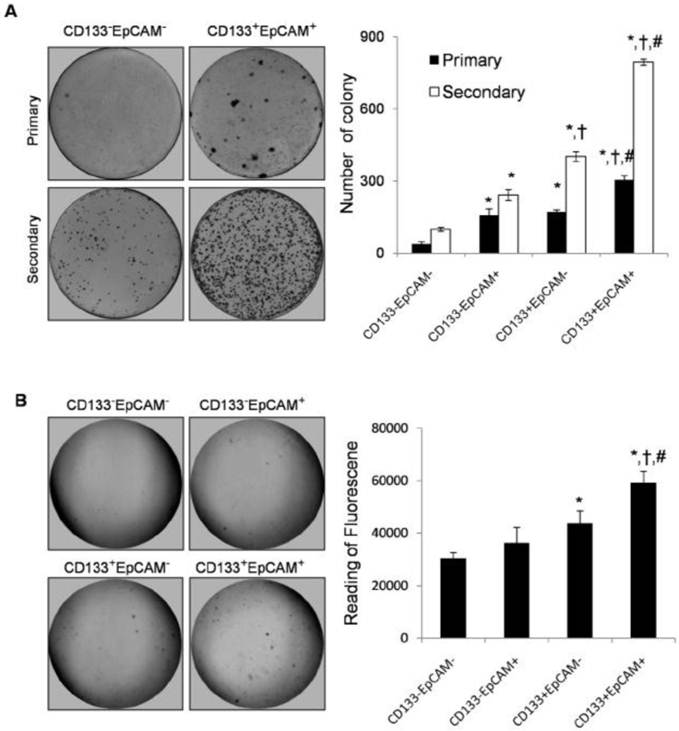
Increase of colony formation ability in CD133+EpCAM+ cells
Plate primary colony formation assay showed that CD133+EpCAM+ cells markedly enhanced bigger and more tumor colonies by 1.8-fold, 1.9-fold and 7.9-fold than CD133+EpCAM-, CD133-EpCAM+ and CD133-EpCAM- cells respectively. Secondary colony formation assay also showed that CD133+EpCAM+ cells formed bigger and more tumor colonies than other three cells (Fig. 3A). In soft agar colony formation assays, CD133+EpCAM+ cells induced bigger and greater numbers of tumor colonies than CD133-EpCAM+, CD133+EpCAM-, CD133-EpCAM- cells. The reading of fluorescence of CD133+EpCAM+ cells was higher than that of other three cells (Fig. 3B). The data suggested that CD133+EpCAM+ cells possessed stronger clonogenic ability than other three cells.
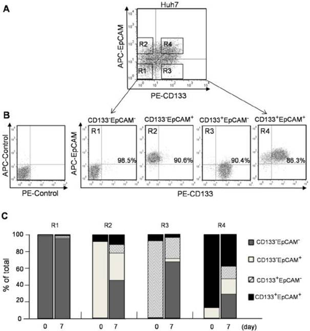
CD133+ and EpCAM+ phenotypes enhanced the differentiation of Huh7 cells. A. CD133-EpCAM- (R1), CD133-EpCAM+ (R2), CD133+EpCAM- (R3) and CD133+EpCAM+ (R4) phenotypes of Huh7 cells were isolated by flow cytometry sorting. B. The sorting purity of R1, R2, R3 and R4 was detected respectively. C. R1, R2, R3 and R4 phenotypes were incubated for 7 days, and analyzed by flow cytometry. Data were from two independent experiments.
 (Click on the image to enlarge.)
Figure 4
Colony formation ability was increased in CD133+EpCAM+ Huh7 cells. A. In plate colony formation assay, various phenotypes were cultured for 14-21 days, and then the primary colonies were replanted for another 14-21 days. Each experiment was performed three times. B. In soft agar colony formation assay, various phenotypes were planted on soft agar and cultured for 8-10 days. The stained colonies were photographed, and measured by the fluorescence reader. Data were from triple separate experiments. *P<0.05 to CD133-EpCAM-, †P<0.05 to CD133-EpCAM+, #P<0.05 to CD133+EpCAM-.
 (Click on the image to enlarge.)
Flow cytometry analysis and sorting
Cells were resuspended in PBS and incubated with FcR blocking reagent (Miltenyi Biotec, Germany) for 10 min. Then cells were stained with the directly conjugated monoclonal antibodies, anti-human CD133-PE, anti-human IgG-PE isotype (Miltenyi Biotec, Germany), anti-human EpCAM-APC, anti-human IgG-APC isotype (R&D, USA) for 30-40 min in 4ºC. IgG isotype control was incubated in parallel. Flow cytometry analysis was performed on Accuri C6 (BD Biosciences, CA) using CFlow (BD Biosciences, CA) software. Cell sorting was performed on BD FACS Aria I (BD Biosciences, CA) using FlowJo (Tree Star, Oregon) software. Sorted cells were cultured in DMEM supplemented with 15% FBS for 7 days, then detected again by flow cytometry.
cell culture
Human hepatocellular carcinoma Huh7 cells were obtained from the ATCC (Frederick, MD). Huh7 cells were cultured in DMEM supplemented with 10% inactivated fetal bovine serum and 1% penicillin-streptomycin (Gibco, USA). Human hepatocellular carcinoma Bel7402, Bel7404 and HepG2 cells were provided by the Cell Bank of Institute for Biological Sciences, China Academy of Sciences (Shanghai, China). Human hepatocellular carcinoma SMMC7721 cell was obtained from Cancer Institute of CAMS (Beijing, China). These four cell lines were cultured in RPMI 1640 medium (Hyclone, UT) supplemented with 10% inactivated fetal bovine serum and 1% penicillin-streptomycin. All cells were incubated at 37ºC with 5% CO2.
4. Langkah-langkah apa yang dilakukan untuk menentukan apakah seseorang kurang pendengaran atau tidak ?
a. Tes suara bisik
Caranya ialah dengan membisikkan kata-kata yang dikenal
penderita dimana kata-kata itu mengandung huruf lunak dan
huruf desis. Lalu diukur berapa meter jarak penderita dengan
pembisiknya sewaktu penderita dapat mengulangi kata-kata yang
dibisikan dengan benar. Pada orang normal dapat mendengar
80% dari kata-kata yang dibisikkan pada jarak 6 s/d 10 meter.
Apabila kurang dari 5 - 6 meter berarti ada kekurang
pendengaran. Apabila penderita tak dapat mendengarkan katakata
dengan huruf lunak, berarti tuli konduksi. Sebaliknya bila
tak dapat mendengar kata-kata dengan huruf desis berarti tuli
persepsi.
Apabila dengan suara bisik sudah tidak dapat mendengar dites
dengan suara konversasi atau percakapan biasa. Orang normal
dapat mendengar suara konversasi pada jarak 200 meter.
b. Tes Garpu Suara
Dengan garpu suara frekuensi 64, 128, 256, 512, 1024, 2048
dan 4096 hz, dibunyikan dengan cara tertentu lalu disuruh
mendengarkan pada orang yang dites. Bila penderita banyak tak
mendengar pada frekuensi rendah berarti tuli konduksi. Bila
banyak tak mendengar pada frekuensi tinggi berarti tuli
persepsi.
Kemudian dengan garpu suara frekuensi 256 atau 512 hz
dilakukan tes-tes Rinne, Weber dan Schwabach sehingga lebih
jelas lagi apakah tuli penderita dibagian konduksi atau persepsi.
c. Tes dengan Audiometer
Hasil dari tes pendengaran dengan audiometer ini digambar
dalam grafik yang disebut audiogram. Apabila pemeriksaan
dengan audiometer ini dilakukan, tes-tes suara bisik dan garpu
suara tak banyak diperlukan lagi, sebab hasil audiogram lebih
lengkap. Dengan audiometer dapat dibuat 2 macam audio-gram :
• Audiogram nada murni (pure tone audiogram)
• Audiogram bicara (speech audiogram)
Dengan audiometer dapat pula dilakukan tes-tes :
• tes SISI (Short Increment Sensitivity Index), tes Fowler
dimana dapat diketahui bahwa kelainan ada di koklear atau
bukan.
• tes Tone Decay dimana dapat diketahui apakah kelainan
dibelakang koklea (retro cochlear) atau bukan. Kelainan retro
coklear ini misalnya ada tumor yang menekan N VIII
Keuntungan pemeriksaan dengan audiometer kecuali dapat
ditentukan dengan lebih tepat lokalisasi kelainan yang menyebabkan
ketulian juga dapat diketahui besarnya ketulian
yang diukur dengan satu db (desibel).
d. Tes dengan "Impedance" meter
Tes ini paling obyektif dari tes-tes yang terdahulu. Tes ini
hanya memerlukan sedikit kooperasi dari penderita sehingga
pada anak-anak di bawah 5 tahun pun dapat dikerjakan dengan
baik. Dengan mengubah-ubah tekanan pada meatus akustikus
ekterna (hang telinga bagian luar) dapat diketahui banyak
tentang keadaan telinga bagian tengah (kavum timpani). Dari
pemeriksaan dengan Impedancemeter dapat diketahui :
• Apakah kendang telinga (membrana timpani) ada lobang
atau tidak.
• Apakah ada cairan (infeksi) di dalam telinga bagian tengah?
• Apakah ada gangguan hubungan antara hidung dan telinga bagian tengah yang melalui tuba Eustachii.
• Apakah ada perlekatan-perlekatan di telinga bagian tengah
akibat suatu radang.
• Apakah rantai tulang-tulang telinga terputus karena kecelakaan
(trauma kepala) atau sebab infeksi.
• Apakah ada penyakit di tulang telirigastapes (otosklerosis).
• Berapa besar tekanan pada telinga bagian tengah.
Kategori
- Masih Kosong
Blogroll
- Masih Kosong