
Pengamatan Jaringan Pada Tumbuhan dan Hewan_Praktikum 8
A. Title
Observation of Networks in Plants and Animals
B. Purpose
1. Knowing Embryonic Tissue in Plants and Animals
2. Differentiate the structure of plant and animal tissue
3. Knowing the Location of the Network
4. Know the Basic Network in Plants and Animals
5. Know the support network in plants and animals
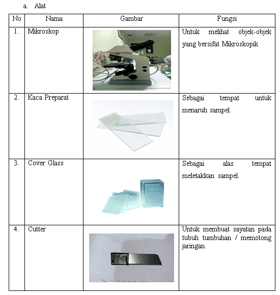
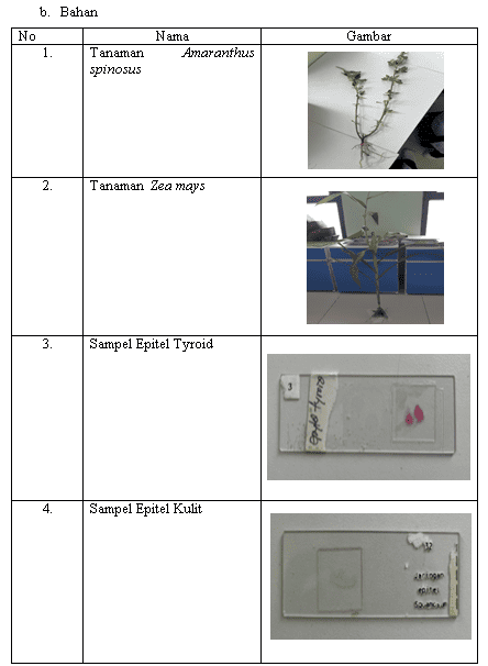
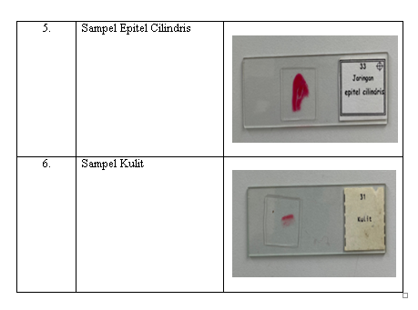
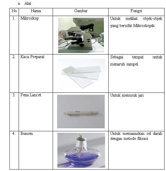
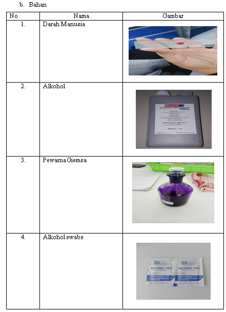
C. Tools and Materials
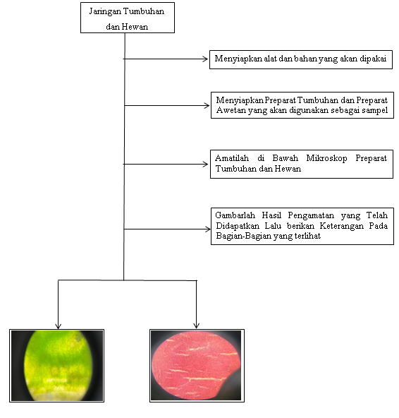
D. Work Procedures
E. Results of Observation
F. Results of Discussion
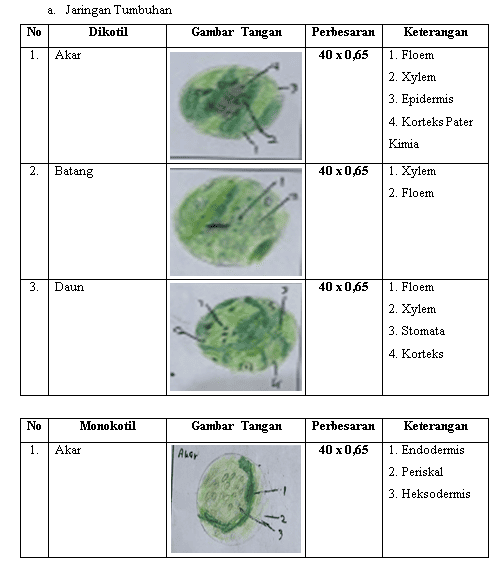
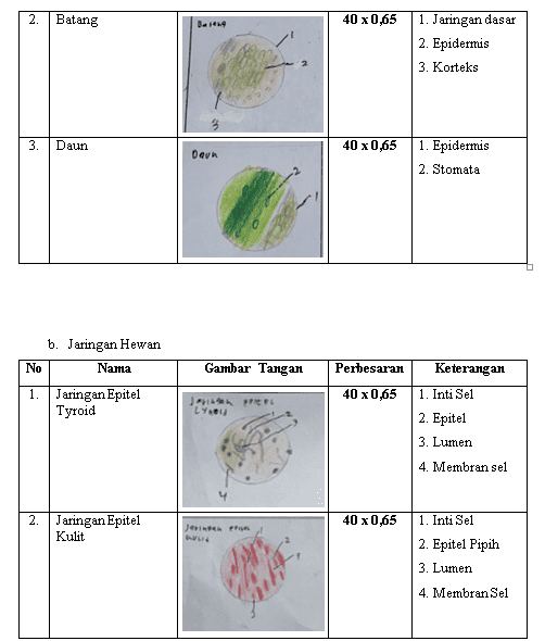
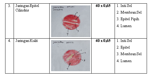
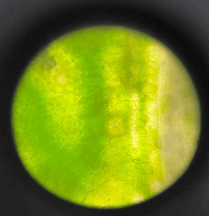
Based on the results of the observations we made, in the first experiment, which was the observation of plant and animal networks, in dicotyledonous plants. Permanent tissue is a tissue composed of adult cells that have been differentiated, but in certain circumstances can be meristematic again. Permanent tissue consists of protective tissue, basic tissue (parenchyma), reinforcing tissue, and transport tissue. For animals, there are two groups of tissue, namely seed tissue and body tissue. The seed network actively divides itself to produce new seeds. Body tissue includes epithelial tissue, connective tissue, muscle tissue, and nerve tissue. In the plant tissues that we observed in this practicum, there are several plant constituent tissues which are protective tissues (epidermis). fruit. In this practicum, almost all parts of the plants we observed have epidermis, only the roots of Amaranthus spinosus have epidermal tissue. Basic tissue (parenchyma), parenchyma tissue is a tissue formed from living cells, with varying morphological and physiological structures and still performing physiological processes. In this practicum, the basic network is only found on corn stalks.
For observations on corn stalks, the parts that have been observed are in accordance with the existing literature, for corn roots, the parts that are in our experiment are lacking for the hairy layer, outer cortex fiber, and pericycle that should be there. For the observation on the stem of Zea mays is in accordance with the available literature. And for the observation of Amaranthus spinosus that can be seen in the observation, we only see the xylem and phloem parts. Because at the time of observation the preparation used broke. But actually on Zea mays. there is a part that shows the network of cortex, epidermis, xylem and phloem. For oryza sativa there are similarities with the literature between the networks, differences
the form of the organizing network. However, there are slight differences due to unclear observation results.
According to (Herliani. & Elsje. T. 2020). Plant tissue on the stem consists of several main layers that support the function and growth of the stem. The outermost part is the epidermis, which protects the stem from physical damage and water loss. Under the epidermis, there is a cortex that plays the role of storing food reserves. The next layer is the vascular network consisting of xylem and phloem. Xylem functions to transport water and minerals from the roots to all parts of the plant, while phloem transports the results of photosynthesis from leaves to the rest of the plant body. Between the xylem and the phloem there is a cambium, a meristematic network that produces new cells so that the stem can grow (secondary thickening).
DOCUMENTATION
PENGAMATAN BENTUK SEL DARAH MANUSIA MELALUI METODE PEWARNAAN GIEMSA_PRAKTIKUM 7
A. Title
Observing the Shape of Human Blood Cells Through the Giemsa Staining Method
B. Purpose
1. Students Skilled in Making Blood Smears That Can Give a Clear Picture of the Forms of Blood Cells
C. Tools and Materials
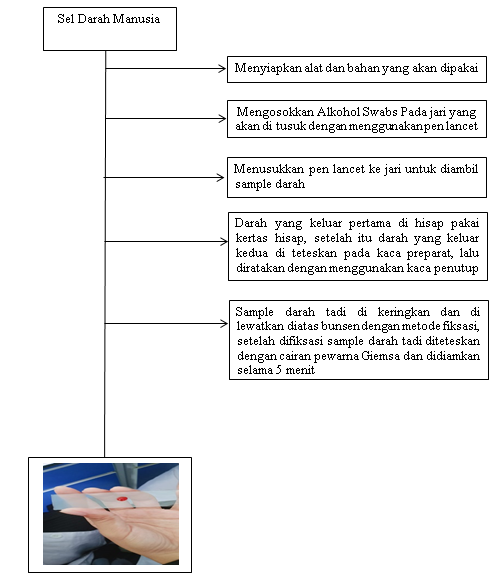
D. Work Procedures
E. Observation Results
F. Discussion Results
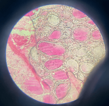
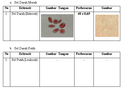
Based on the results of the observations made by the practitioner, in the first experiment, which is the observation of the shape of human blood cells through the gieamsa staining method using a 40 × 0.65 lens, the practitioner did not find results because the microscope was problematic so the practitioner did not find a clear picture. Then in the second observation result with the same lens, which is 40 × 0.65, the practitioner still did not find the result because there was too much Giemsa dripping. In the third observation with the 100 × 1.25 lens, the practitioner did not find results because the smear process was not perfect so it was difficult to get the blood cell type. However, in the observation of the fourth experiment with a 40 × 0.65 lens, the practitioner found the result of finding an image of red blood cells. After finding red blood cells, the practitioner still tried to get white blood cells but the practitioner did not find results so in this observation the practitioner only found one type of blood cell namely red blood cells.
DOCUMENTATION
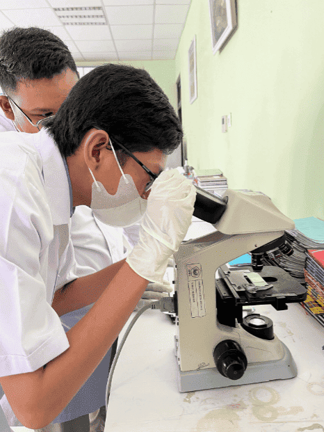
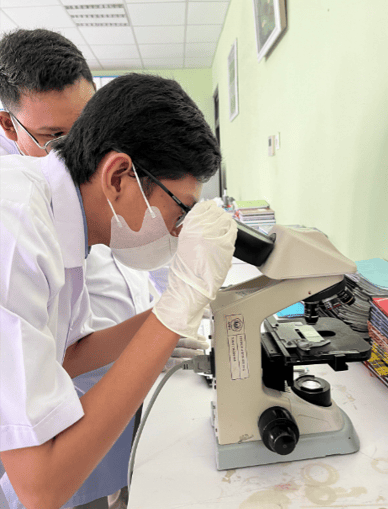
Observation Under the Microscope
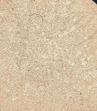
Red Blood Ladybug
Blogroll
- Masih Kosong